|
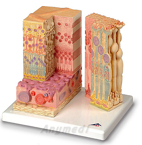
눈의 조직 모형
이 모형은 맥락막과 공막을 가진 망막의 구조를 나타낸다.
왼쪽 부분은 모형 옆쪽 층에 있는 공막의 부분과 관다발층이 포함 되어있는 망막의 완전한 구조를 보여준다
오른쪽 부분은 모형의 부분적인 확대모습을 보여주며 광수용기와 색칠된 세포의 구조들을 보여준다.
왼쪽부분은 850배 확대, 오른쪽부분은 3800배 확대된 모습이다.
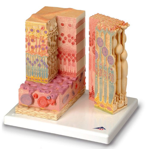
3B MICROanatomy™ Eye
The MICROanatomy™ Eye model illustrates the microscopic anatomical structure of the retina with choroid and sclera.
The left block-like, layered side of the eye model shows the complete structure of the retina including the supplying vascular layer and parts of the sclera from a light microscopic view.
The right part of the eye model is a sectional enlargement.
MICROanatomy™ Eye shows the microscopic structure of the photoreceptors and the cells of the pigmented layer.
Left part of MICROanatomy™ Eye 850-times enlarged - right part 3800-times enlarged. You've never seen the human eye like this before!
25 x 23 x 18.5 : 1.2 kg
 | 
; "확대")