- 현재 위치
- home > 인체모형/실습모형/CPR > 두개골모형/해골모형 > [3B] 7분리 고급형 두개골모형 (A283)
; "확대")


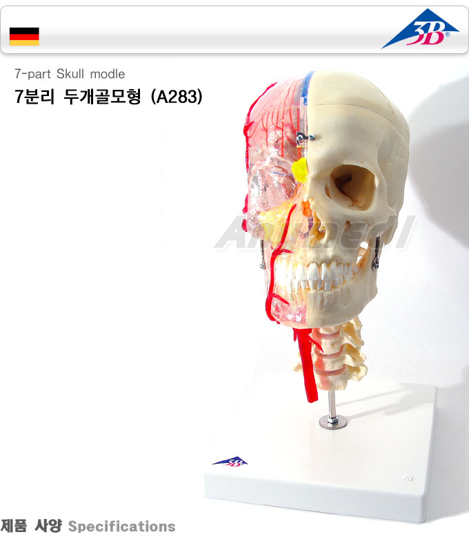
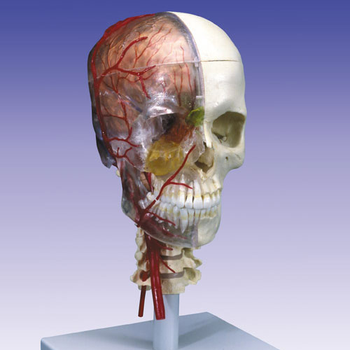
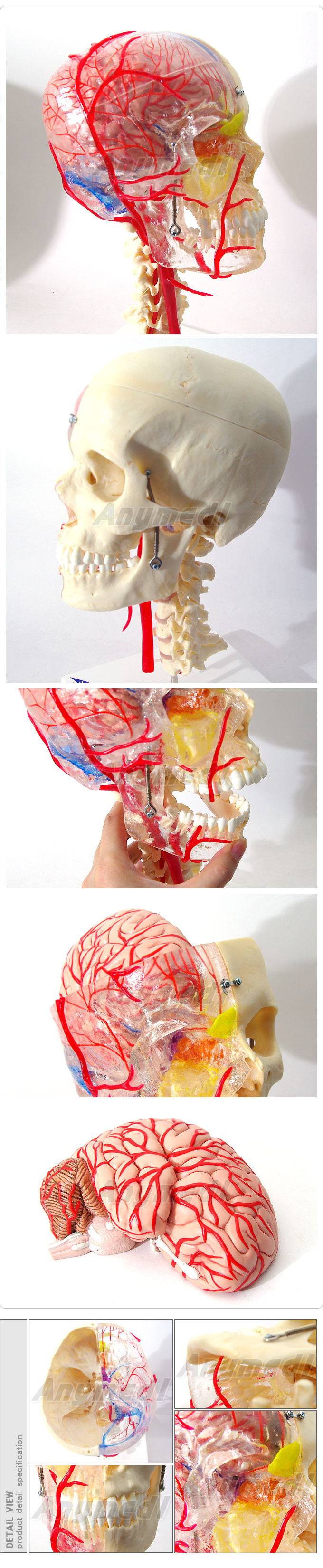
두개골 모형 7조각으로 분리되는 모형
1.고급형 교육용 두개골 모형이며 7부분으로 나뉨
2.투명한 반 두개골은 각기 다른 색으로 두개골,골공,골열구 등을 표시하며 특히 뇌수막 동맥 혈관과 뇌경막을 붉은색과
푸른색으로 연결
3.다른 한면은 두개관을 통해 뇌 위치,골공 위치를 표시할 수 있음
4.투명한 치아구조와 턱 구조 비격막과 하악골 등의 관찰 가능
5.턱 아래 부분은 유연한 구조로 척추와 연결
3B Scientific System Skull-Didactic Deluxe Skull, 7-part
This worldwide unique and high-quaity skull will leave no questions in the study of anatomy unanswered!
The possibility to transfer the structures visible on the transparent half to the bony half give this skull a special didactic balue.
ON the right, transparent skull half the paranasal sinuses can be easily located even from the outside, since these are marked
in different colors: maxillary sinus(yellow), ethmoidal cells(orange), frontal sinus(green), sphenoidal sinus(purple).
The cranial sinuses and the neck and facial arteries are also shown in color: sinuses of dura mater(blue), common carotid
artery, external and internal carotid artery and the branches of the meningeal artery(red).
One brain half, which is also visible through the skullcap, visualizes the brain position and the course of the sinuses.
The peridontal pockets and dental roots can be studied through the transparent jaw.
The lower jaw is mounted flexibly to demonstrate the masticatory movements.
The skull is mounted on the cervical spine and can be disassembled into both halves of the skullcap, the left half of the base
of skull, the nasal septum, the complete mandible and brain half.
35 x 18 x 18 cm; 1.0 kg
|
■ 상품기본정보 |
| ||||||||||||


 |