- 현재 위치
- home > 인체모형/실습모형/CPR > 손목관절/어깨관절/무릎관절 > [3B] 신장결석모형 K29

; "확대")


신장 결석 모형
위 제품은 신장 결석에 관한 환자와 요로 결석증이 있는 환자에 대한 도움을 주는 모델입니다.
우측 신장이 열려진 상태의 실물크기입니다.
신장 캘러시즈와 신장 골반 및 요관에서 일반적으로 발견되는 위치에서 결석을 확인할 수 있습니다.
신장 피라미드 부분
캘릭스그룹 위쪽의 시작되는 부분
신장 외피 부분
캘릭스그룹에 연결되는 혈관 부분
요관 부분 4가지 색깔은 다양한 신장 결석을 보여줍니다.
Kidney Stone Model
This model is a helpful tool to inform patients about kidney stones (nephrolithiasis) and urinary stones (urolithiasis).
It shows an opened right kidney in natural size.
The renal calices, the renal pelvis and the ureter are opened as well so that concretions or stones can be identified in the
following typical positions:
* In the area of the renal pyramids
* In the area of origin of the upper calix group
* In the renal cortex
* In the connecting tubule of the lower calix group, causing congestion of the minor calices (partially closed, partially opened)
* In the ureter 4 original color pictures on the base show various kidney stones.
19 x10 x16.5 cm: 0.18 kg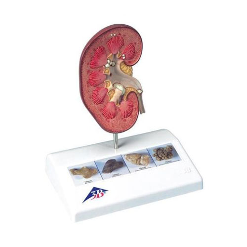
| ■ 상품기본정보 |
| ||||||||||||


 |